Inside Chameleon Skin: The Science of Color Change
Start inside a single reflective cell. Imagine plates of guanine—small enough to earn the name nanocrystals—stacked with a rhythm. That rhythm is a lattice. Change the spacing of the rhythm and you change which colors of light bounce back toward the world. That is the core of the modern scientific picture of dramatic chameleon color change: not a brush dipping into new paint, but a living optical device retuning itself. Pigments still sit in the audience and on stage with it. Temperature-relevant reflectors wait deeper in the skin. This article begins at the lattice, zooms outward, and ends with methods and open questions.
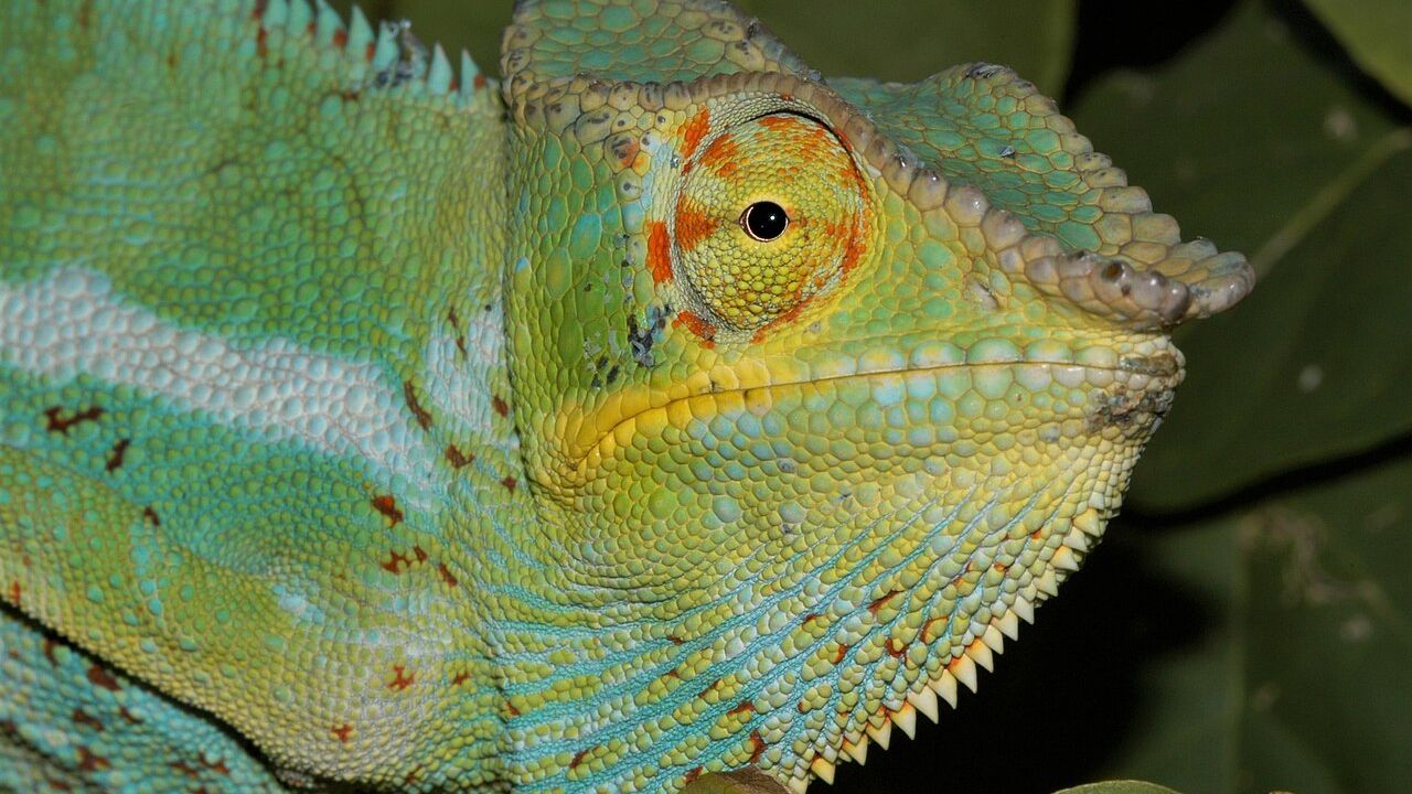
Close view of textured skin for educational context
Meet the nanocrystal lattice
Iridophores are skin cells specialized for reflection. In the superficial iridophore layer of panther chameleons, guanine nanocrystals form an ordered array. Order matters. Periodic structure plus contrast in refractive index yields selective reflection—behavior familiar from engineered photonic crystals and from other animals’ structural colors.
Structural-color takeaway
Inside chameleon skin, the headline scientific story is structural color under nervous and hormonal control: a guanine nanocrystal lattice that expands and contracts to rewrite reflected light, pigments that tint and melanin that darkens, and deeper crystals that reflect near-infrared for thermal physics—demonstrated most clearly in panther chameleons, still incomplete in cellular control, and not automatically identical in every species.
Remember the hinge: spacing changes color; physiology changes spacing; pigments finish the picture; heat-relevant layers work in parallel; open questions remain. That is the science inside the skin.
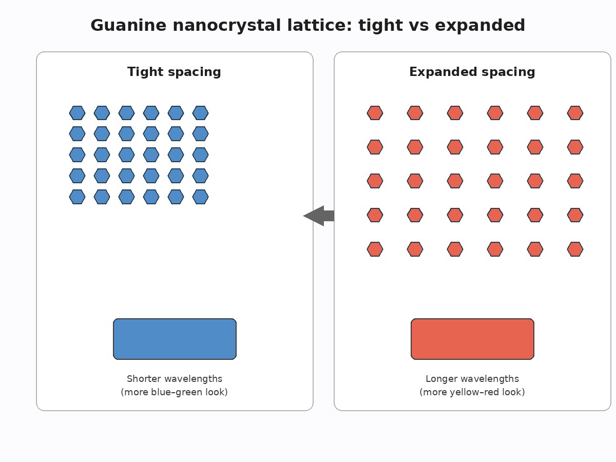
Before-and-after spacing in a nanocrystal lattice
Calling the array a lattice is not poetic license. Imaging shows geometric organization; optical models predict reflectance from that geometry; measured skin spectra move when geometry moves. The 2015 Nature Communications study by Teyssier and colleagues made that chain of evidence central for panther chameleons. Inside the cell, color change is first a materials-science story: spacing as a control parameter.
Guanine is a familiar biological molecule (one of the DNA bases), here deployed as a crystalline reflector rather than as genetic code. Evolution reused chemistry for optics.
Meeting the lattice first also resets intuition. People often begin with the whole animal—“green lizard becomes angry red lizard”—and work backward. Starting in the iridophore makes the causal arrow clearer: geometry changes, spectrum changes, appearance changes. Behavior and hormones sit upstream of geometry; pigments sit alongside it. The nanocrystal array is the hinge.
How spacing turns blue into yellow or red
Photonic reflection depends on characteristic distances in the lattice relative to the wavelengths of light. When nanocrystals sit closer together, the skin’s selective reflection tends to favor shorter wavelengths—feeding blue-leaning appearances once the rest of the tissue is accounted for. When the animal’s physiology drives the lattice to expand, the preferred reflected band slides toward longer wavelengths. Yellow, orange, and red displays become available without manufacturing a new pigment library on the spot.
Think of a tunable filter built from spacing rather than from dye concentration. The animal’s calm-to-excited transition, in optical terms, is often a compact-to-expanded transition in the superficial lattice. Overlying and neighboring pigments then remix that reflected light into the greens, golds, and crimsons observers name in the field.
Speed follows from mechanism: rearranging spacing is faster than synthesizing saturating amounts of new pigment. That is why scientific accounts of rapid change lean on structure first.
It helps to separate three related words. Hue shifts when the reflected wavelength band moves. Brightness shifts when overall reflectance or melanin influence changes. Saturation shifts when spectra become more or less peaky relative to broadband reflection. Lattice expansion is especially powerful for hue; melanin is especially powerful for brightness; pigments influence both hue and saturation. Inside the skin, those knobs can turn together or somewhat apart.
Pigments still matter (the supporting cast)
Structural color does not exile pigment biology. Chromatophores with yellow and red pigments absorb and transmit selectively, staining the structural base note. Without them, many “classic” chameleon greens and warm display tones would not appear as they do. Melanophores deploy melanin to darken. Dispersed or optically emphatic melanin lowers brightness and can flatten perceived chroma; reduced melanin influence lets photonic colors flare.
A rigorous scientific description therefore refuses a false choice—pigment or structure. The accurate frame is interaction: lattice sets a shifting reflectance peak; pigments and melanin sculpt the spectrum that exits. Educational shortcuts that say “only crystals” or “only paint” both fail the skin’s actual stack.
Historically, chromatophore language dominated because pigments are chemically intuitive and histologically familiar. Structural color demands comfort with wave optics. Once that comfort arrives, chameleon skin becomes a teaching model for biophotonics as much as for herpetology: a vertebrate tissue that actively tunes a photonic crystal while still using classical pigments.
Deep cells that bounce infrared
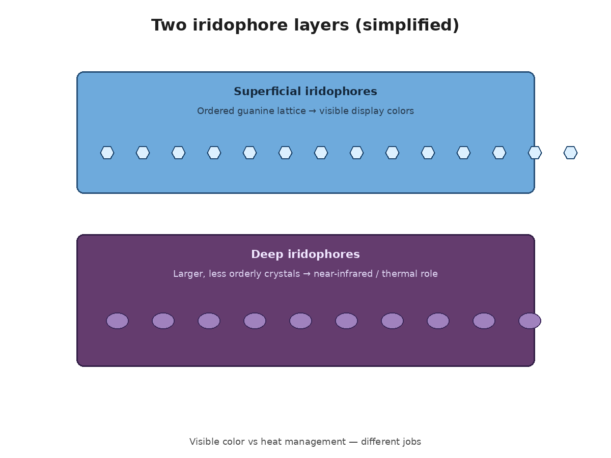
Below the showy superficial lattice lies another iridophore population described in panther chameleons: larger crystals, less orderly packing, strong reflectance in the near-infrared. Human eyes do not paint pictures from that band the way they do from yellow and blue, but solar energy cares. Reflecting near-infrared can alter heat gain.
Researchers present this deeper layer as a candidate for passive thermal roles—a structural contribution to thermoregulation distinct from the social billboard above. Inside chameleon skin, then, “optics” splits: visible signaling versus heat-relevant bounce. Both are science of color and reflectance; only one dominates Instagram.
The deep-layer story also shows why “color change” is a slightly narrow English phrase. Near-infrared reflectance is not a color humans name, yet it is part of the same reflective tissue system. Scientific writing sometimes speaks of spectral management or reflectance change to keep that broader physics in view.
How researchers figured this out (plain English)
The panther chameleon breakthrough was not a single magic photograph. It was a combination of tools:
Microscopy to visualize guanine nanocrystal organization in iridophores from animals in different physiological states (for example, calmer versus excited).
Spectroscopy to measure which wavelengths the skin actually reflected in those states.
Physical modeling of photonic lattices to test whether measured spacings predict measured colors.
Comparison across conditions to show that lattice expansion tracked the shift toward longer-wavelength reflectance.
University-facing explanations of the work (including those connected with the Geneva research community involved) stressed the take-home: chameleons actively tune a photonic crystal in their skin. For readers asking how chameleons change color scientifically, that methods trio—see the crystals, measure the light, model the physics—is the template.
No need to overclaim. The paper did not claim to have finished the physiology of control, and it did not sample every chameleon species on the planet.
Plain-English methods matter for public trust. When readers hear “scientists discovered nanocrystals,” they may picture a single eureka glance. The real path is slower: prepare tissue, image structure, measure light, build a model, test whether the model’s predictions match the spectra, compare physiological states. That loop is how structural-color claims earn their keep.
Superficial display layer and deeper infrared-reflective layer
Open questions and careful limits
Control signals inside the cell
What molecular actors stretch or compact the lattice? Which receptors, ions, cytoskeletal elements, or second messengers couple nerve and hormone signals to nanocrystal spacing? The 2015 authors noted that mechanisms remained to be determined. Educational writing should repeat that humility. Filling the gap with invented pathways would be storytelling, not science communication.
Until control is mapped in detail, we can still say that physiology drives spacing and that spacing drives color—while admitting how the cell’s machinery executes the resize is incomplete.
That gap is normal in integrative biology. Organismal function and tissue optics can be solid while cell-molecular actuators remain partly unknown. Good science communication names the gap instead of papering it with speculative diagrams presented as fact.
Not every species is a panther chameleon
Panther chameleons (Furcifer pardalis) earned center stage because their displays are extreme and their skin was measured carefully. Other species may share motifs—iridophores, pigments, melanin—yet differ in lattice order, tuning range, speed, or the balance of social versus thermal versus cryptic functions. Camouflage’s contribution, likewise, is not a constant across the family; it varies by species and ecological context.
Scientific generalization should be labeled as such. Mechanism demonstrated in one star species is a hypothesis generator for others, not an automatic fact about “the chameleon.”
Camouflage’s variable contribution belongs in the same caution bin. Some contexts and species may lean more on crypsis; flashy social change in display specialists still aligns poorly with a camouflage-first story. Writers should keep that nuance next to the photonic mechanism so mechanism and function do not get flattened into one cartoon.
Additional open areas include development (how juveniles build lattices), pathology and nutrition effects on structural color, and the neural circuits that time social displays. Those frontiers keep the topic alive beyond a single landmark paper.